
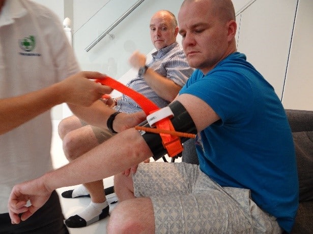
My brush with Cancer
Last year I went to see my GP as I felt something was pulling inside my right arm. The doctor looked and said it wasn’t anything to worry about. Later this year having returned unwell from nursing in Africa, I managed to see another GP. I asked her to have a look at my arm and she immediately referred me to Guys Dermatology. Two weeks later I was seen by the Dermatologist, they scanned me all over with their microscopic lights and said they weren’t overly worried but would like me to have a biopsy. A month later and I had a biopsy. A few weeks after that and I was told that they had removed a large, deep, advanced stage 3a malignant melanoma. I had gone to the appointment on my own as I really wasn’t expecting this diagnosis and it was all a bit of a shock.
This summer was then dominated by various scans in order to try and establish how far it might have spread and the likely prognosis. Initially the outlook was pretty grim and scary.
Care and support at Guy’s
The Consultants at Guys were amazing. The Thursday clinic consists of oncologists, dermatologists and plastic surgeons, they work as an incredible team and you really do feel you are in the best possible hands. You are allocated a cancer nurse specialist and encouraged to attend future appointments with your family. I was given a leaflet of key contacts and was called and offered emotional and financial support if I needed it. Everyone is as kind and supportive as possible, but it doesn’t stop it from being frightening and they will give you the worst scenarios, which is a pretty bleak picture. Be prepared to re-evaluate your life. It makes you think very deeply and consider if you really have left the legacy you would be proud of. It prompts deep conversations and brings you face to face with your mortality.
Gradually over the next few months, I had an MRI scan of my brain and PET scan of my whole body to search for any other tumours over 5mm in size.
My tumour was atypical as it was within my arm, and the dermis (top layer of skin) was not involved. Usually, malignant melanomas result from moles or other surface damage to the skin. Because there was no surface element to my tumour, the one they have found is not likely to be the primary. They may never locate the primary, so need to keep scanning me – just in case!
Further surgery and lymph node check
Fortunately, the MRI and PET scans did not reveal any metastasis or spread. Consequently, they decided that I just needed further surgery to take a 2cm margin around the original tumour site. They therefore removed the size of a tennis ball from my right upper arm (the original tumour was about the size of a golf ball). They also suggested that I might like to get my sentinel lymph node removed. The sentinel node is the first node that drains the lymph from around the site of the tumour and so this would be the first one to be affected if the cancer had spread to my lymph.
Thankfully the cancer doesn’t appear to have spread to my lymph or anywhere else. However, because they can’t find the primary, they are kindly following me up every 3 months and scanning me regularly too.
Why I’m sharing my story
Everything is healed, I am fit and well and I am sharing this as I hope some of the things I have experienced and learned about might be helpful to others on a similar journey.
There is information about scans and procedures that I wish I had known prior to attending. It would have made some of my experiences a little less intimidating and helped me know what to expect.
Key advice:
Check yourself and ask someone else to check for you too.
Some usefull websites to help you know what to look for:
Check moles regularly – you are looking for moles that bleed, have changed recently increased in size, changed in shape, deepened in colour, itch, bleed or have any surrounding inflammation.
Take photos and measure moles you are monitoring. Some people even get professionally mole mapped!
Visit your GP if you are worried about anything and if you are not happy with the response ask for a referral to Dermatology.
Check strange lumps and bumps – mine wasn’t a mole.
Ask for a referral to a specialist if you are worried – if my GP had referred me a year ago, everything would have been caught earlier and would not have been nearly so scary.
Prevention:
They don’t know exactly what causes melanoma but believe it may have a strong link with UV exposure, particular as a child. Sunbeds are also implicated.
Wear sun cream, cover up and look at the UV index. I wasn’t looking at the UV index at all – it is on the weather forecast and on the weather apps. It tells you when the UV is at its absolute highest and this varies considerably according to where in the world you are and how cloudy it is too.
Preparation for appointments:
It can all appear very scary. You will receive information piece by piece and it can be overwhelming. Ask them to clarify if you don’t understand anything and take someone with you – ideally ask them to take notes. If you don’t ask questions, the team will assume you have understood everything.
Keep notes of any questions that come to mind and don’t worry about asking them all. If it is on your mind then clearly it is important to you.
You will be given details of nurse specialists who you can contact. Do not be scared to contact them, they are there to help answer your questions and put your mind at ease through the scary process.
Sometimes doctors and nurses use medical terminology, without meaning to and this can be confusing and a bit intimidating. Just ask them to explain in simple terms, they won’t mind at all.
PET scan
What is a PET scan?
A PET scan is a test that creates 3 dimensional (3D) pictures of the inside of your body. PET stands for positron emission tomography.
A PET scan uses a mildly radioactive liquid called a radioactive tracer. It shows areas of your body where cells are more active than normal. It’s used to help diagnose some conditions, including cancer. It can help to find out:
- where the cancer is
- the size of the cancer
- whether the cancer has spread
Where you have your PET scan
You usually have a PET scan in the radiology or nuclear medicine department as an outpatient. These scanners tend to be only in the major cancer hospitals. So you might have to travel to another hospital to have one.
How long PET scans take
A radiographer operates the scanner. The scan usually takes between 20 and 30 minutes. But you will be in the department for around 2 hours. This is because you need to arrive about an hour before the scan to have the injection of the radioactive tracer. Your appointment letters will usually tell you how long your appointment will be.
PET scans are often combined with CT scans to produce more detailed images. These are called PET-CT scans. You can also have an MRI scan with a PET scan. These are called PET-MRI scans.
If you need a PET scan, you will be in a specialist department.
Getting ready for your appointment
Remove jewellery prior to your appointment and leave it at home.
Wear clothing without metal fastenings. If possible wear a sports bra, that way, you may be able to keep it on.
When you arrive at the department
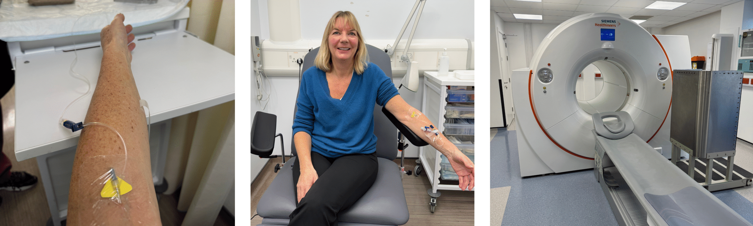
You will sit in the waiting room and then get taken through by a nurse or radiographer, who will check your name and date of birth and ensure you are booked for the right procedure. They will then put a cannula into your hand or arm. This is for the radioactive isotope, that will be injected exactly an hour before your scan. This will enable the machine to scan and identify any tumours, or other areas of interest larger than 5mm.
You will be then taken through to the ‘hot’ waiting area. Where you will be able to lie on a trolley, or sit on a chair. You will be waiting for at least an hour. Take something to read, or do, or simply have a snooze. This area has high levels of radioactivity and so you will not be allowed visitors and the staff will be restricting their time in there as much as possible. Exactly an hour before your scan, a nurse or radiographer will come and inject the isotope into your cannula. This does not hurt and you should not feel anything at all.
They will then remove the cannula and leave you to allow the isotope to travel round your bloodstream. Just before the hour is up, the tannoy will call your name and ask you to go to the loo to pass urine before your procedure. They will double check that you have removed all jewellery and have no metal fastenings. I was wearing jeans and so unfortunately had the indignity of being scanned with my jeans round my knees!!! I’ll know next time and hence I’m sharing the information with you too.
Having your PET scan
The scan itself is quite chilly. You will be asked to lie on the bed on the machine and this bed will move into the machine. It is not as claustrophobic as an MRI scanner, a bit larger and very quiet. The scan itself is relatively quick and not uncomfortable. If you do feel claustrophobic in any way, practise deep breathing and grounding exercises. You will have a panic button just in case, but you shouldn’t need it. If you do have any concerns, speak to the radiographer before your procedure. Your results will be through in a week or so and you will discuss them with the doctor at your next appointment.
MRI scans
An MRI is a type of scan that creates pictures using magnetism and radio waves. MRI scans produce pictures from angles all around the body and shows up soft tissues very clearly.
MRI stands for magnetic resonance imaging. They take between 15 and 90 minutes.
A radiographer operates the scanner and looks after you while you have your scan.
Why might you be referred for an MRI scan?
An MRI scan can be used to look at most areas of the body and is used to:
- find a tumour
- find out how big it is and whether it has spread (stage a cancer)
- measure blood flow
- to check how well treatment is working
You might have an injection of a special dye called a contrast medium before the scan. This is to help make the scan pictures clearer.
MRI scans are particularly good for:
- brain tumours
- primary bone tumours
- soft tissue sarcomas
- tumours affecting the spinal cord
- tumours in the pelvic organs, including prostate, bladder, womb and ovarian tumours
MRI scans are also done in a specialist department, although my latest one at Thomas’s will be done in a portacabin in the car park! My first one was at Guys and was done on a Sunday afternoon when everything else in radiology was shut!
Remove all jewellery prior to your appointment and leave it at home. Although they did say my gold wedding ring would have been okay. Wear clothing without metal fastenings and a sports bra, not an underwired or adjustable one.
If you are prone to claustrophobia, talk to your GP in advance of the scan and if very concerned that you won’t cope in the scanner, ask them to prescribe some Lorazepam or some other short term sedative to take just before the scan. If you have taken a sedative it would be sensible to have a friend or family escort you home from your appointment.
MRI – of the brain, noisy and unpleasant
Use deep breathing, grounding techniques, it is claustrophobic and a bit frightening and you are in there for quite a while.
Cold – take a jumper
Don’t wear jeans or anything with a metal fastening to your MRI or Pet scan – if you have trousers without a metal fastening, then you can wear them during the scan – depending where you are being scanned and whether you need a contrast medium.
Wear a sports bra without any metal fixings and then you can keep it on.
You may still be asked to wear a hospital gown. If you are cold, do ask for a blanket. The scanning suites are usually quite chilly.
Sentinel node scan
A sentinel lymph node biopsy (SLNB) is a way of checking the lymph nodes closest to the melanoma. It is usually an optional procedure, and you may be told about this test even if your lymph nodes do not look or feel swollen. It is an incredibly sensitive test and can help to find very small amounts of melanoma that have spread to the lymph nodes.
The sentinel node is the first node lymph fluid drains to from the melanoma. If the melanoma has spread, the sentinel node is the most likely to be affected. There may be more than 1 sentinel node and they are likely to remove all those that they can see are affected.
How does the test work?
The doctors will inject a radioactive isotope to identify which nodes are affected. The blue cobalt dye can stay in the skin for many months after the procedure.
This test is not suitable for everyone. Your doctor or specialist nurse may talk to you about having an SLNB if the melanoma is: 1mm or thicker thinner than 1mm, but there are other risk factors.
You usually have an SLNB at the same time as surgery to remove a surrounding area of healthy tissue from around the original melanoma location (called wide local excision).
An SNLB provides more accurate information about the stage of the melanoma and the risk of the melanoma coming back.
Your doctor will ask you to decide whether to have an SLNB. They will explain the possible advantages and disadvantages. It may help you and your doctor to plan the best treatment for you. If you decide not to have an SLNB, you still have the wide local excision.
If there are no melanoma cells in the sentinel lymph nodes, it is unlikely the melanoma has spread to other lymph nodes. You will not need further tests or treatment.
Advice for your wide excision and sentinel lymph node biopsy
They will usually ask you to get to your appointment early. You will probably need to go to the day surgery, or admissions unit first, where they will prepare you for your later surgery – height, weight, BP, temperature… explain your procedure and consent you for the operation.
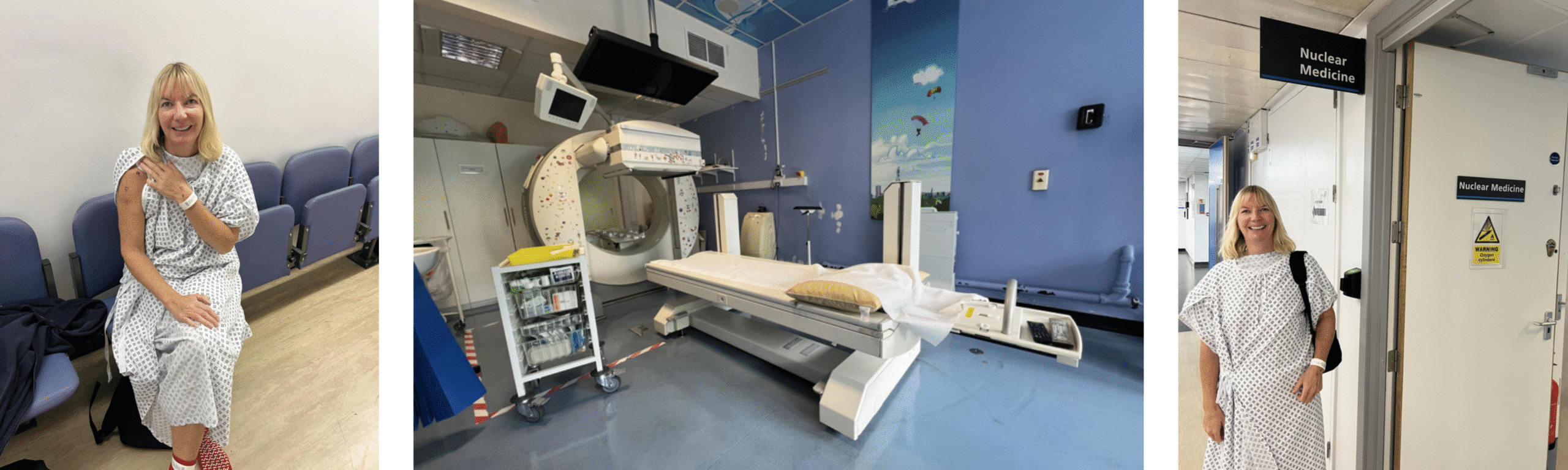
You will then go up to nuclear medicine to be injected with the isotope. The scanning room is very cool, so if you are in a theatre gown, don’t get cold and ask for a blanket or cover if you are feeling chilly.
Having the injections and first scan
You will lie on the table and they will inject 4 corners of your biopsy scar with the isotope. It is very slim needles and so is not painful. They will then cover the injection site and push the table you are lying on, into the scanner. To scan your lymph nodes, they will put arm supports and ask you to have your arms outstretched either side so they can scan your armpit.
You will be under the scanner for up to an hour with this initial scan. They will need you to wriggle out if your gown slightly to expose your armpit and they will then use a cobalt pen to mark any lymph nodes they have found and take still photos. Then they will move the scanner to 90 degrees and take photos and images from the side for about 20 minutes. They will also hold a screen from the other side so that they can see an outline of your body on the scan, to help the surgeon further contextualise where they need to operate.
The whole procedure is uncomfortable, but not painful. You will be lying still on a hard scanning platform for a long time, with your arms outstretched and your arms may get pins and needles! I also got absolutely freezing!!
The isotopes are technetium-99m injection and a cobalt pen to mark and screen to see body outline.
Surgery
As soon as you have been scanned you will go down to day surgery theatre to have your surgery. You will be prioritised as you will be a ‘hot’ radioactive patient and they will want to get you into theatre as quickly as possible whilst the isotopes are still at their maximum intensity so they can scan in theatre and see which of your lymph nodes are affected. The plastic surgeons will then remove these at the same time as undertaking the wide excision.
Once you have had your surgery, you will wake up in recovery and then go through to the patient collection area. There you will be offered a drink and something to eat and allowed to go home once they are sure you are not bleeding and have managed to have a pee. You will need to be collected by someone else
Post surgery
Closely follow all the directions from your doctors and nurses. The team kindly prescribed me some Fucidin H so that I could administer to myself if my wound was showing any early signs of infection.
Once the wound is healed and the stitches have either dissolved or been removed, then you can start using Vaseline or Bio oil. Massaging this in certainly reduced my scarring.
About us
First Aid for Life offer award-winning first aid training tailored to your needs. Our trainers are highly experienced medical, health and emergency services professionals. They will tailor the training to your needs. Courses for groups or individuals at our venue or yours.
We strongly advise that you attend a fully regulated Practical or Online First Aid course to understand what to do in a medical emergency. In addition, please visit First Aid for Life or call 0208 675 4036 for more information about our courses.
First Aid for life provides this information for guidance and it is not in any way a substitute for medical advice. First Aid for Life is not responsible or liable for any diagnosis made, or actions taken based on this information.


Such a brave person. Incredible strength is needed at times like this and I have so much admiration for you and for what you have been through AND for sharing.
Keep well?
Thank you so very much – all good now x
Thank you for your excellent explanation. It is so helpful for anyone who is going through a cancer diagnosis.
Thank you – thinking of everyone going through this. It is scary!
My last comment should not have had a question mark but a smile! Keep well?
Thank you for sharing Emma, and being helpful, even when YOU are a patient. Wishing you a full recovery.
Thank you so much. So glad it was helpful, I thought long and hard whether it was a good thing to share. However, I think knowing what to expect always make things a bit less frightening.
Aww Emma, wishing you all the best for the future, may that evil thing never return, and thank you for sharing your story!
Thank you so much. I am glad it was helpful to share. Thinking of all the people out there processing such news, it is frightening, but hopefully this might help in some very small way xxx
I am so sorry to hear you have been through this Emma but I am very happy to hear the outcome has been so positive.
Thank you for sharing with everyone, these stories are so helpful and it takes courage to do this.
Stay well and take care. xxx
Thank you so much Henrietta. You never know whether it is the right or wrong thing to do and if it is over-sharing! But if it helps someone, then it has been the right decision! Hope all is well with you Emma
I am so glad to hear you are well. Thank you for sharing this in such a clear and accessible and positive way.
Thank you so much, I really appreciate your message